ERCP系列二:插管篇

 游小土等 2人推荐
ERCP系列二:插管篇
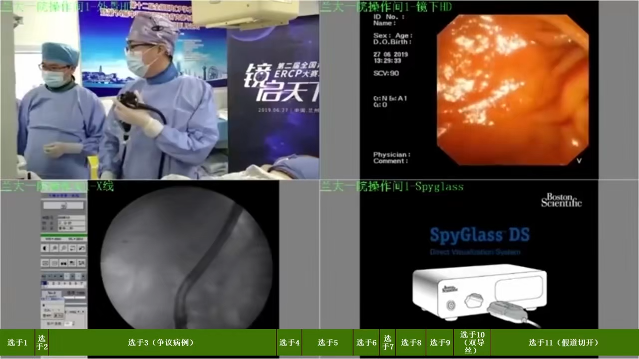
ERCP插镜视频(镜启天下全国ERCP大赛A组决赛选手视频):


- 寻找乳头及开口
拉直内镜后,即可在十二指肠降段沿纵形皱襞走向寻找乳头。乳头部基本结构如图1-48所示。副乳头通常位于主乳头右上方,相距约20mm左右,较主乳头为小,通常无缠头皱襞。

发现乳头后还必须辨清开口,以便插管。乳头开口形态一般分为五型,如图1-49所示:①绒毛型:与乳头外观一致,由较粗的绒毛组成,开口不明显;②颗粒型:开口部绒毛粗大,活动较频繁,常有色调改变;③裂口型:开口呈裂口状;④纵口型:开口呈纵线状裂形开口,有时呈条沟样;⑤单孔型:开口部呈小孔状,硬而固定。


- 主乳头插管方法
患者取俯卧位,在插管及注射造影剂之前,常规摄右上腹部平片,观察有无钙化及气泡,乳头插管最好在平面中心位置进行,造影剂充填导管以排出导管内气泡,但勿将造影剂排至十二指肠内,因十二指肠内造影剂过多会刺激肠蠕动,影响插管。
借助于内镜弯曲部的上下左右弯曲功能以及旋转镜身、使用抬举器,精细调节造影导管的头端;并吸除肠腔内过多气体,推进或回拉内镜,以帮助内镜头端接近乳头(图1-50)。

胰管插管多选择垂直于十二指肠壁,或在1~2点位置进行(图1-51)。避免过度挤压乳头,以免乳头水肿影响插管。插管不顺利时,可多次轻微改变方向和位置。胆管插管多从乳头下方插入,有时通过用导管挑起乳头顶部,向11~12点方向插管 (图1-52)。多数初学者认为胰管插管易于胆管插管。


Illustration of typical anatomy of the ampulla of Vater. A short common channel (CC) is present. The septum (S) separates the common bile duct (CBD) and pancreatic duct (PD).
Vater壶腹的典型解剖图:存在短共同通道(CC)。隔膜(S)分离胆总管(CBD)和胰管(PD)。

A Illustration of separate openings for the CBD and PD. B Actual papilla with separate openings. Bile can be seen around the biliary opening
A:胆总管和胰管单独开口示意图。B:胆管胰管分别开口实际乳头示意图,胆道开口周围可见胆汁。

Illustration of the location of the CBD opening on the papilla when viewed en face.
从正面看时,胆总管开口在乳头上的位置示意图。

A Cannulation of the bile duct using a standard catheter. Note steep trajectory that the biliary axis takes. B Bowed sphincterotome facilitates biliary cannulation by achieving the steep axis.
A:使用造影导管胆管插管,注意胆道轴的陡峭轨迹。B:弓形刀通过拉弓实现陡峭的轴线有利于胆道插管

Biliary cannulation using a guidewire(使用导丝进行胆道插管)

A The tip of a sphincterotome has passed from the papillary orifi ce through the intraduodenal portion of the bile duct during attempted biliary cannulation. B After severing the intraduodenal portion.
A:尝试胆管插管时,弓形刀从十二指肠乳头一侧穿出。B:行假道切开
- 视频:


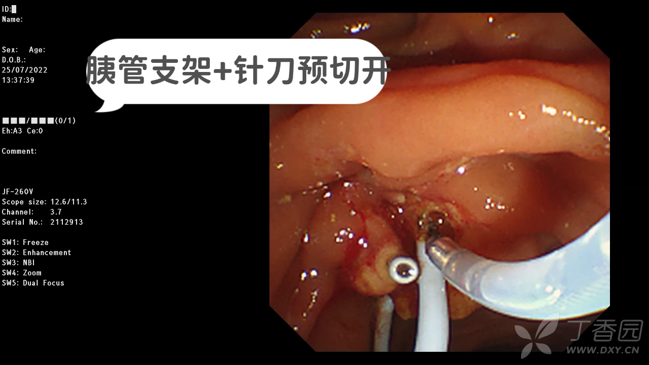
A Endoscopic view of biliary cannulation above the previously placed pancreatic duct stent. B Fluoroscopic view of the same patient. Pancreatic duct stent is seen (arrow).
A:胰管支架置入后再选择性胆管插管。B:同一病人X线图示,箭头指胰管支架

A Illustration of double wire technique. B Endoscopic view of sphincterotome alongside pancreatic guidewire. C Fluoroscopic view of double wire technique.
A:双导丝插管示意图。B:双导丝法插管内镜示意图。C:双导丝法插管X线示意图
- 视频:
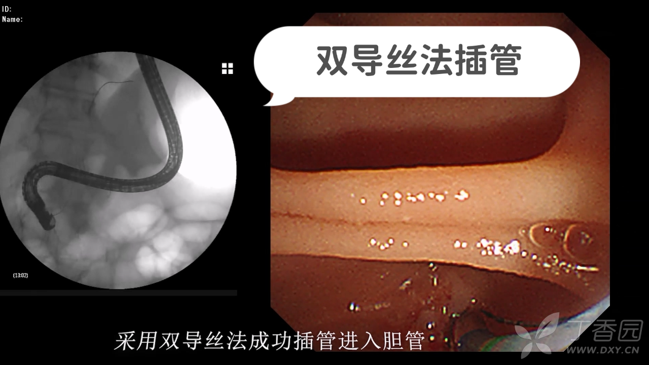

Illustration of precut sphincterotomy technique using a needle-knife beginning at the papillary orifi ce. A Without pancreatic duct stent. B With pancreatic
duct stent.针状刀预切开技术。A:无胰管支架。B:有胰管支架
- 视频:

A Illustration of precut fi stulotomy technique. B Successful precut fi stulotomy. The cut
was performed with a needle-knife and is well above the papillary orifi ce (arrow).
A:针状刀乳头开窗示意图。B:十二指肠乳头上方使用针状刀开窗

Illustration of transpancreatic septotomy technique
经胰管预切开(胆胰隔膜切开)

- 乳头插管困难时的操作流程
ERCP插管视频(镜启天下全国ERCP大赛A组决赛选手视频):