


stranding, a smoky appearance?
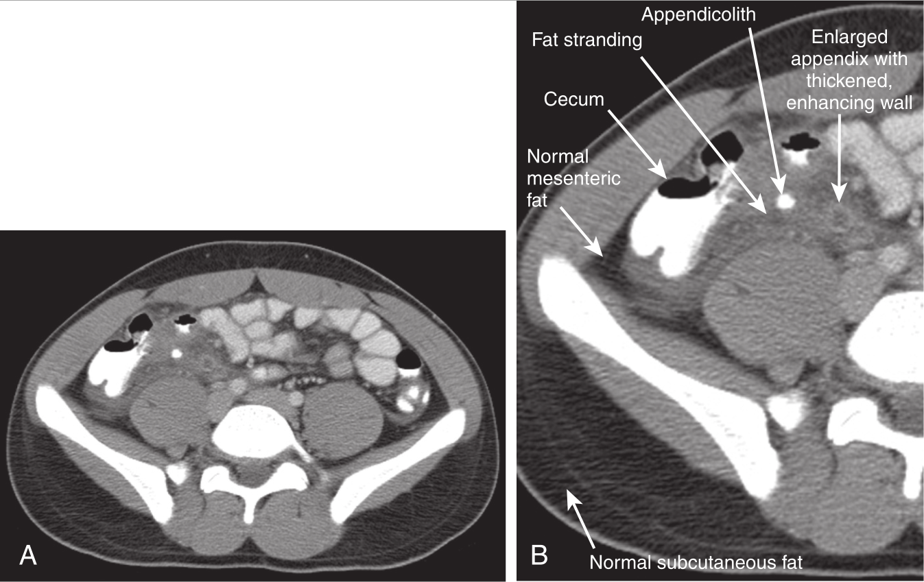
FIG. E1 Appendicitis, computed tomography (CT) with intravenous and oral contrast. This CT demonstrates classic findings of appendicitis in an 18-year-old male with right lower quadrant pain, as seen with CT with IV and oral contrast. Studies suggest that CT without contrast has similar sensitivity and specificity. An enlarged appendix is seen near the cecum as a right lower quadrant tubular structure in short-axis cross section, giving it a circular appearance. The surrounding fat shows stranding, a smoky appearance indicating inflammation (compare with normal mesenteric and subcutaneous fat, which is nearly black). The appendiceal wall shows enhancement, a brightening after administration of IV contrast. This slice also shows an appendicolith, an occasional finding of appendicitis. It does not appear to be within the appendix in this slice, because the appendix bends in and out of the plane of this slice. An appendicolith usually appears as a calcified (white) rounded structure, visible without any contrast. A, Axial CT image. B, Close-up.
图E1:阑尾炎,静脉和口服造影剂 CT。这张CT显示了一名18岁男性右下腹疼痛阑尾炎的典型表现,如静脉注射和口服造影剂的CT所示。研究表明,不加造影剂的CT具有相似的敏感性和特异性。盲肠附近可见增大的阑尾,在短轴横切面上显示为右下象限的管状结构,呈圆形。周围脂肪堆积、烟熏状,提示炎症(与这些几乎是黑色的正常的肠系膜和皮下脂肪相比)。阑尾管壁增强,静脉注射造影剂后变亮。此切片还显示阑尾结石,结石偶尔会发现于阑尾炎。在这个切面中,结石看起来并不在阑尾腔内,因为阑尾并不在这个切面内。阑尾结石通常表现为钙化的(白色)圆形结构,未用造影剂时看不见。A,轴位CT图像。B,静脉期(闭合期)。
stranding, a smoky appearance和Close-up该如何理解?谢谢
最后编辑于 2022-10-09 · 浏览 963


